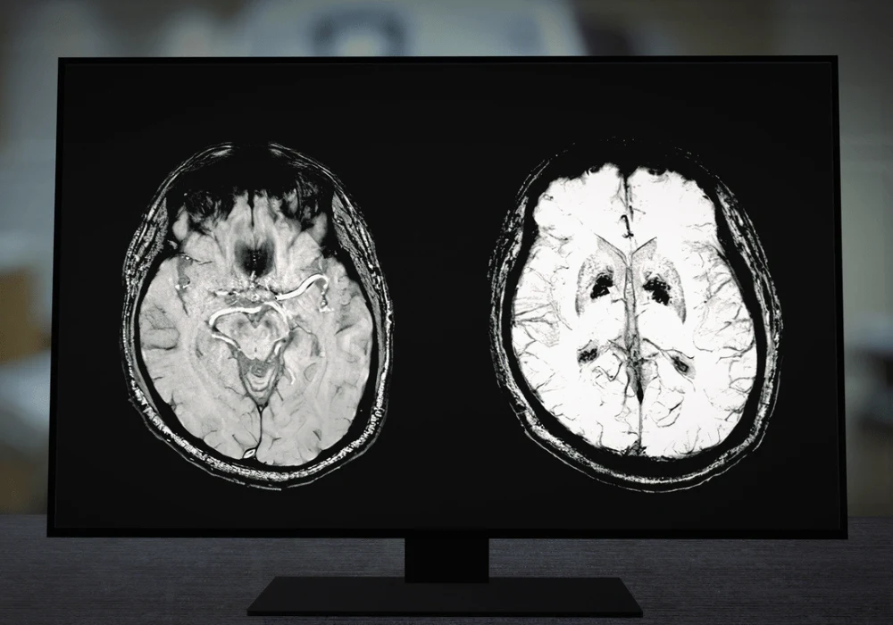
Brain MRI scans are essential for diagnosing a wide array of neurological conditions. However, analysing these images requires substantial expertise, and even experienced radiologists can face challenges in detecting subtle lesions due to the complexity of brain structures. This complexity has prompted the integration of deep learning (DL) models to assist in lesion detection. A recent study reviewed in Radiology has taken these advancements further by incorporating textual data from radiology reports into the DL models to enhance the accuracy and interpretability of brain MRI lesion detection.
Role of Radiology Reports in Enhancing Lesion Detection
One of the study's primary innovations is the integration of radiology reports into deep learning models. Radiology reports offer detailed descriptions of lesion characteristics such as size, location, and signal intensity, which are not always easily detectable in MRI scans. These reports reflect expert knowledge, making them a valuable source of weak annotations for guiding AI systems.
In the study, the DL model “ReportGuidedNet” was trained using textual features extracted from radiology reports to guide its attention toward brain lesion characteristics. This approach eliminated the need for manual image segmentation, which can be labour-intensive and inconsistent. By leveraging radiology reports, the model could focus on critical regions in the MRI images, thus improving lesion detection accuracy. The study demonstrated that this integration outperformed models trained purely on image data, indicating the potential of multimodal approaches combining visual and textual information.
Comparative Performance of ReportGuidedNet and PlainNet
The study compared the performance of two models: ReportGuidedNet, which incorporated textual features from radiology reports, and PlainNet, which relied solely on image data. The results showed that ReportGuidedNet consistently outperformed PlainNet across multiple diagnostic categories. The area under the receiver operating characteristic curve (AUC) for ReportGuidedNet was significantly higher in internal and external test datasets. This improvement was particularly notable in the external dataset, where the performance difference between the two models was more pronounced.
The ability of ReportGuidedNet to generalise across different datasets suggests that incorporating radiology reports enhances the model's robustness. Unlike PlainNet, which struggled with variations in imaging conditions across different centres, ReportGuidedNet maintained stable performance. This robustness is essential in clinical settings where MRI acquisition protocols and scanner parameters can vary widely.
Interpretability and Clinical Utility of Attention Maps
Another crucial aspect of the study was the focus on model interpretability. The main challenge in deep learning for medical imaging is the "black box" nature of the models, where it is difficult to understand how the model arrives at its predictions. To address this, the authors incorporated attention maps into ReportGuidedNet. These maps highlight the regions of the MRI scan that the model focused on when making its predictions, providing radiologists with interpretable visual cues.
The study found that the attention maps generated by ReportGuidedNet were more accurate and clinically relevant than those produced by PlainNet. In particular, ReportGuidedNet was better at localising lesions, even when the lesions were small or subtle. This improved localisation can aid radiologists in making more accurate diagnoses and reduces the risk of missed lesions. The attention maps offer an additional layer of transparency, enabling radiologists to validate the model’s predictions and incorporate their expert judgment into the final diagnosis.
Integrating radiology report-derived textual features into deep learning models significantly advances brain MRI lesion detection. The study demonstrated that ReportGuidedNet, which uses both image and textual data, outperformed traditional models relying solely on images. This multimodal approach improves diagnostic accuracy and enhances the interpretability of the model’s decisions, making it a valuable tool for radiologists. Such innovations will likely play an increasingly important role in medical imaging, leading to more accurate, efficient, and interpretable diagnostic systems.
Source: Radiology
Image Credit: iStock


